The novel diagrams represent a new method
The novel diagrams represent a new method for new students
to learn histology step by step. The novel diagrams are beatiful diagrams that
clarifying the accurate structure of tissues by the microscope in simple very
clear method. Also, they identify the scientific structure of tissues typically
like that by the microscope in simple and very fine method that any one can
learn drawing from them. The novel digrams also depict an imagination that the
micrographs of histology are following the same idea of preparing the sample
and sectioning as showen in the novel diagrams. This helps the new students to
imagin and understand the origen of the micrographs of histology. Also the
diagrammed region of tissue section showing the whole structure of the organ in
one figure with magnification as by the microscope while the microscope field
can,t reveals the whole structure in the same field. The following are
forexamples of novel diagrams:
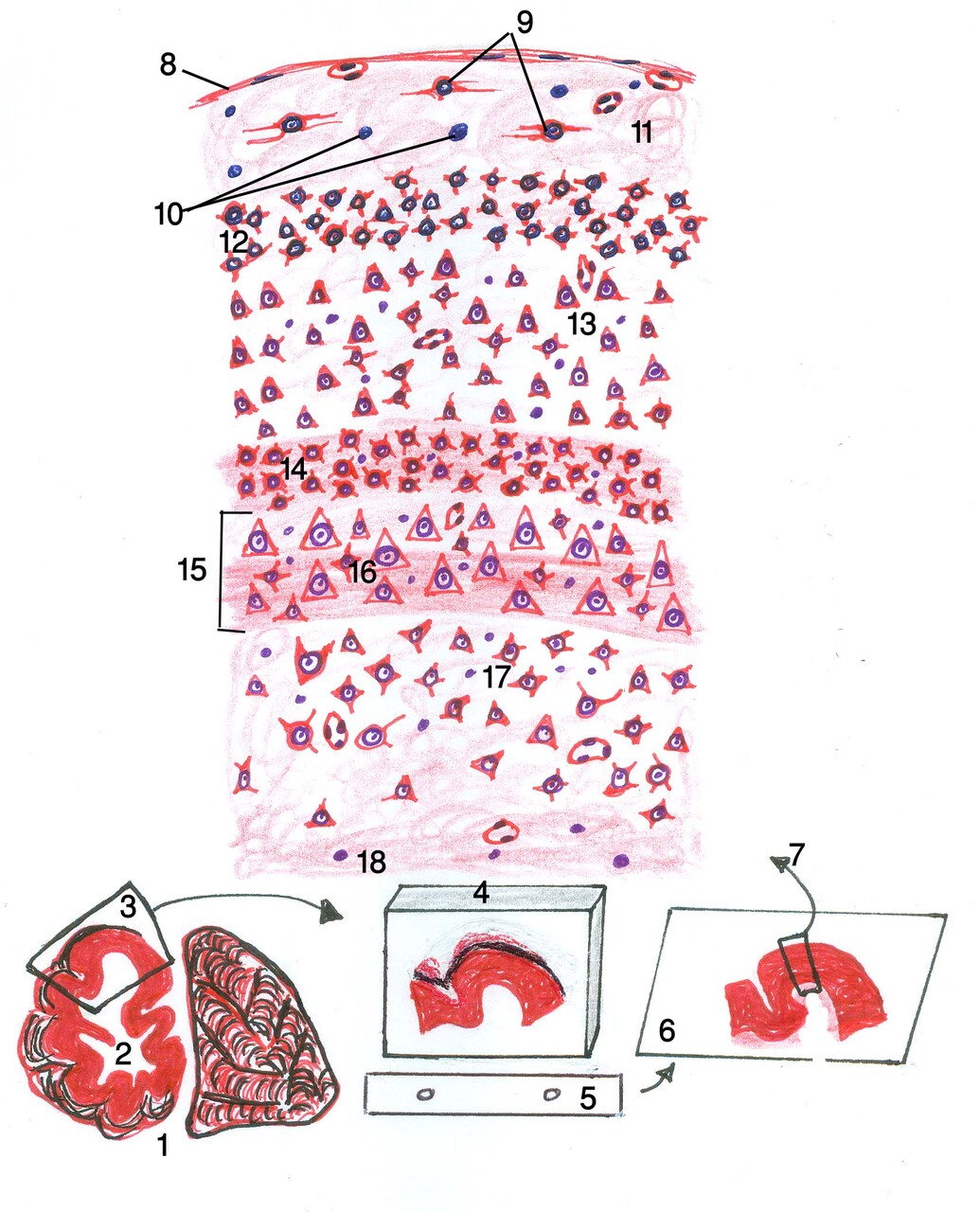
Fig.9: Diagram of cerebrum.
1 : bisection of cerebral hemisphere 2: lateral ventricle 3: sampling 4:
paraffin block containing cerebral sample after processing in position like
that of the microtome 5: microtome knife 6: slide containing stained cerebral
tissue section 7: diagrammed region of tissue section (showing the whole
structure of cerebrum with magnification as by the microscope; while the
microscope field can,t reveals the whole structure in the same field) 8: Pia
mater 9: neurons 10: neuroglia 11: molecular layer 12: external granular layer
13: external pyramidal layer 14: internal granular layer showing external band
of Baillarger 15: internal pyramidal layer 16: internal band of Baillarger 17:
multiform layer 18: medulla.
Post Views : 11