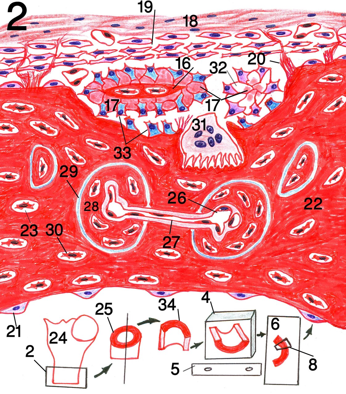
The novel diagrams of my book
The novel diagrams of my book represent a new method for new
students to learn histology step by step. The novel diagrams are beatiful
diagrams that clarifying the accurate structure of tissues by the microscope in
simple very clear method. Also, they identify the scientific structure of
tissues typically like that by the microscope in simple and very fine method
that any one can learn drawing from them. The novel digrams also depict an
imagination that the micrographs of histology are following the same idea of
preparing the sample and sectioning as showen in the novel diagrams. This helps
the new students to imagin and understand the origen of the micrographs of
histology. Also the diagrammed region of tissue section showing the whole
structure of the organ in one figure with magnification as by the microscope
while the microscope field can,t reveals the whole structure in the same field.
Diagram of compact bone (example of novel diagrams).2: sampling 4: paraffin
blocks containing the samples after processing in position like that of the
microtome 5: microtome knife 6: slide containing stained tissue section 8:
diagrammed region of tissue section (showing the whole structure of bone but
with magnification as by the microscope while the microscope field can,t
reveals the whole structure in the same field)
16: calcified matrix 17: secreted uncalcified matrix 18: outer fibrous layer of
periosteum 19: inner osteogenic layer 20: Sharpey,s fibers 21: inactive
osteoblasts of endosteum 22: calcified bone 23: osteocyte lacuna 24: proximal
part of femur bone 25: part from femur shaft 26: blood vessel in Haversian
canal 27: blood vessel in Volkmann’s canal 28: Haversian system 29: cement
substance 30: osteocyte 31: osteoclast 32: differentiation of osteoblasts in
groups that start to secrete bone matrix 33: active osteoblasts 34: sample of
bone from femur shaft.
Post Views : 12