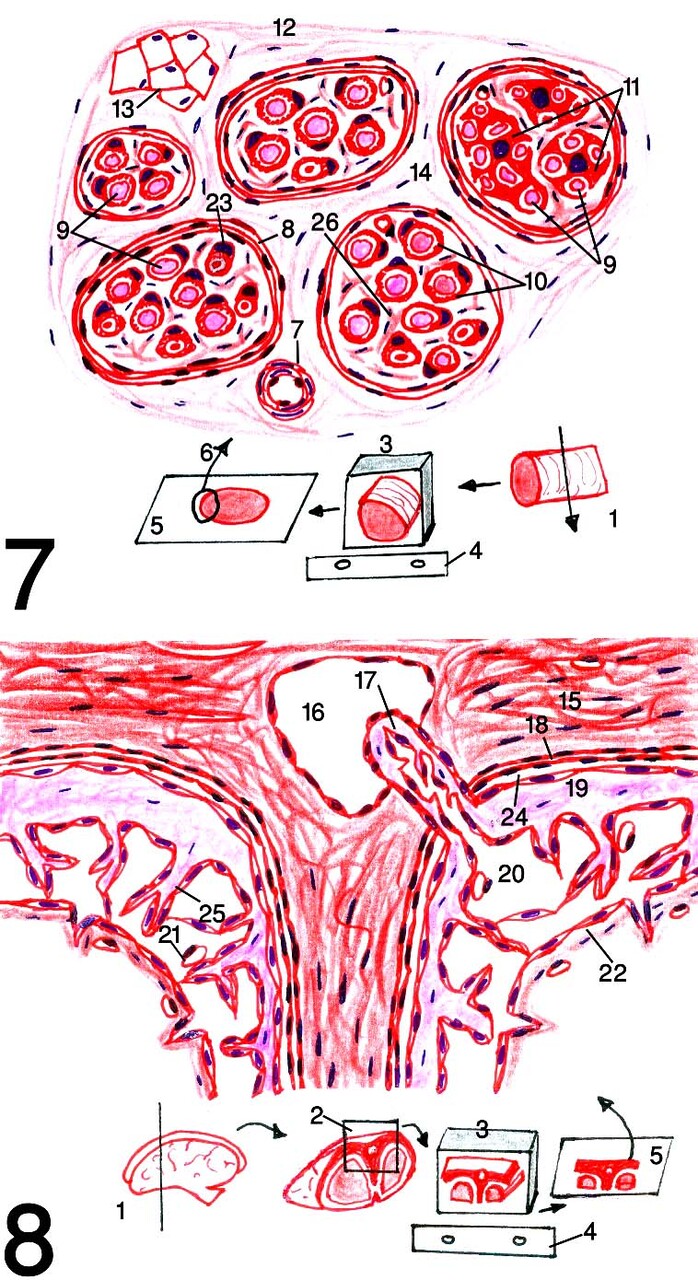
Fig.7: Diagram of nerve.
Fig.8: Diagram of meninges.
1: cutting for sampling 2: identification of meninges sample 3: paraffin blocks containing the samples after processing in position like that of the microtome 4: microtome knife 5: slide containing stained tissue section 6: diagrammed region of tissue section (showing the whole structure of nerve where the whole tissue section of meninges is drawn with magnification as by the microscope because the microscope field can,t reveals the whole structure in the same field) 7: arteriole 8: perineurium 9: nerve fibers or neuron axons 10: myelin sheath 11: Schwann cells of unmyelinated nerve fascicle 12: epineurium 13: adipose tissue 14: connective tissue extension of epineurium 15: thick connective tissue layer of Dura mater 16: venous sinus 17: arachnoid villus 18: condensed fibroblasts of Dura mater 19: arachnoid 20: subarachnoid space 21: blood vessel 22: Pia mater 23: Schwann cell of myelinated nerve fascicles 24: subdural space 25: arachnoid trabeculae 26: endoneurium.
Post Views : 9