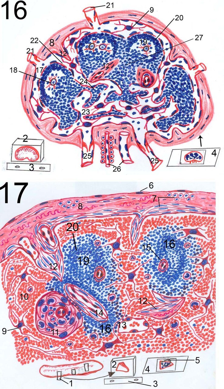
Fig.16: Diagram of lymph node.
Fig.17: Diagram of spleen.
1: sampling of spleen from possible several regions 2: paraffin blocks
containing sample of spleen but containing whole small lymph node after
processing in position as in the microtome 3: microtome knife 4: slide
containing stained tissue section 5: diagramed region 6: visceral peritoneum 7:
smooth muscle 8: capsule 9: reticular cells 10: red pulp 11: ellipsoid 12:
trabeculae 13: splenic sinus 14: white pulp arteriole 15: follicular arteriole
16: white pulp (PALS and lymph nodule) 17: subcapsular sinus 18: dendritic cell
19: germinal center of lymph nodule 20: peripheral dark zone of lymph nodule
21: afferent lymphatic vessels 22: trabecular sinus 23: medullary sinus 24:
medullary cords 25: efferent lymphatic vessels 26: blood supply of lymph node
27: diffuse lymphatic tissue.
Post Views : 10