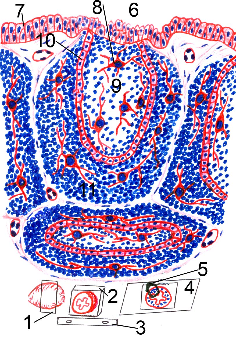
Fig.11: Diagram of bursa Fabricius.
1: sampling from bursa Fabricius
2: paraffin block containing the bursa sample after processing in position like that of the microtome
3: microtome knife
4: slide containing stained tissue section of the bursa
5: diagrammed region of the bursa section
6: lumen of the bursa
7: pseudostratified columnar epithelium
8: reticular epithelial cell
9: medulla of lymphoid follicle
10: cuboidal cells
11: cortex of lymphoid follicle.
Post Views : 13