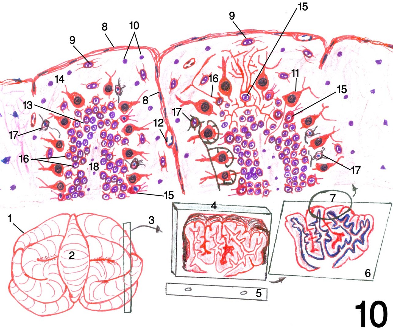
Fig.10: Diagram of cerebellum.
1: cerebellum in simple gross anatomy 2: vermis 3: sampling
4: paraffin block containing cerebellum sample after processing in position
like that of the microtome 5: microtome knife 6: slide containing stained
cerebellum tissue section 7: diagrammed region of tissue section (showing the
whole structure of cerebellum with magnification as by the microscope; while
the microscope field can,t reveals the whole structure in the same field) 8:
Pia mater 9: small stellate neurons 10: neuroglia 11: Purkinje cells forming
piriform layer 12: blood vessel 13: granular cell layer 14: molecular layer 15:
Golgi cells 16: granule cells 17: basket cells 18: medulla.
Post Views : 19