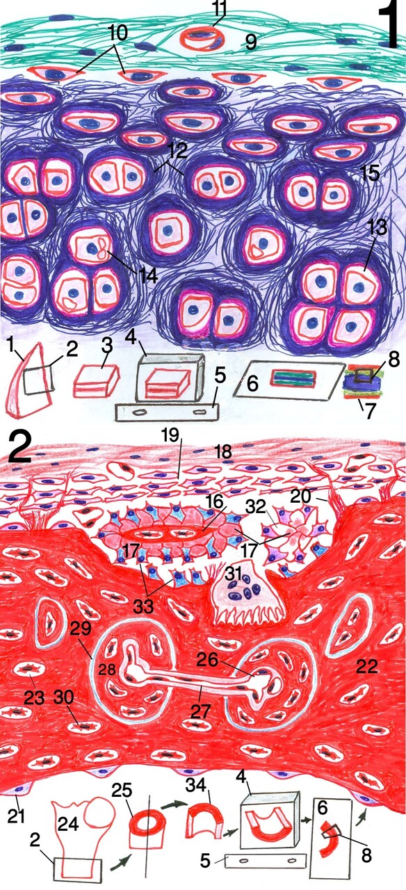
Fig.1: Diagram of elastic cartilage, (Weigert,s elastic & Light green).
Fig.2: Diagram of compact bone.
1: ear pinna 2: sampling 3: sample from ear pinna 4: paraffin blocks containing the samples after processing in position like that of the microtome 5: microtome knife 6: slide containing stained tissue section 7: skin of ear 8: diagrammed region of tissue section (showing the whole structure of both elastic cartilage and bone but with magnification as by the microscope while the microscope field can,t reveals the whole structure in the same field) 9: outer fibrous layer of perichondrium 10: chondroblasts of inner chondrogenic layer 11: blood vessel 12: territorial matrix 13: chondrocyte lacuna 14: chondrocyte containing vacuoles 15: elastic fibers 16: calcified matrix 17: secreted uncalcified matrix 18: outer fibrous layer of periosteum 19: inner osteogenic layer 20: Sharpey,s fibers 21: inactive osteoblasts of endosteum 22: calcified bone 23: osteocyte lacuna 24: proximal part of femur bone 25: part from femur shaft 26: blood vessel in Haversian canal 27: blood vessel in Volkmann’s canal 28: Haversian system 29: cement substance 30: osteocyte 31: osteoclast 32: differentiation of osteoblasts in groups that start to secrete bone matrix 33: active osteoblasts 34: sample of bone from femur shaft.
Post Views : 15